
Figure 4. Cartilage-specific β-CATENIN signaling regulates SOC formation.
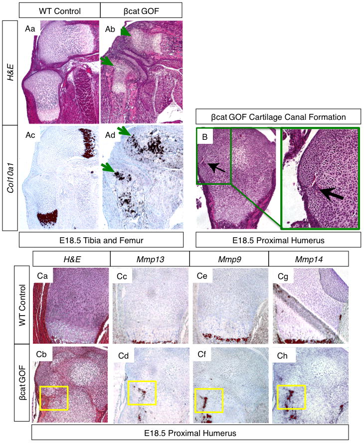
(A) H&E stained sections of E18.5 tibia and femur from WT (Aa) and βcat GOF (Ab) littermates, green arrows depict hypertrophic chondrocytes in the epiphyses of βcat GOF specimens (5x). In situ hybridization for Col10a1, a hypertrophic chondrocyte marker, in adjacent WT (Ac) and GOF (Ad) sections. Green arrows highlight hypertrophic marker expression in the epiphyses of βcat GOF hind limb sections. (B) H&E stained sections at 10X and 20X of the proximal humerus from an E18.5 βcat GOF embryo. Green box and black arrows highlight a forming cartilage canal. (C) H&E stained sections (Ca–b) of WT and βcat GOF proximal humerus with corresponding in situ hybridization of adjacent sections for Mmp13 (Cc–d), Mmp9 (Ce–f), and Mmp14 (Cg–h). Yellow boxes highlight an area of cartilage canal formation (10X).