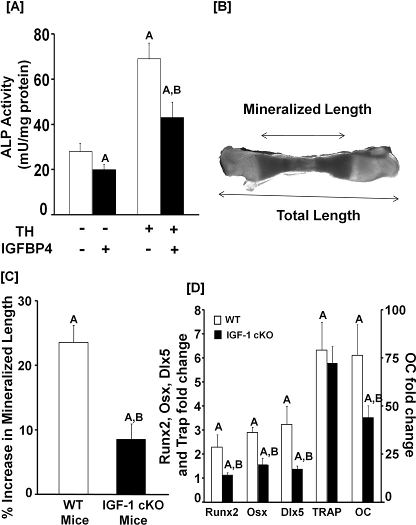
Figure 7. TH biological effects in bone cells is in part mediated via local IGF-I.
[A]: Neutralization of IGF-I with inhibitory IGFBP-4 blocks the TH-induced increase in ALP activity. MC3T3-E1 cells were incubated with or without T3 or IGFBP-4. Three days later, cells were lysed for ALP activity determinations and total protein measurements. Values are the Mean ± SEM (n = 8). A = P < 0.01 vs. vehicle control. B = P < 0.01 vs. T3. [B]: A microscope image of a mineralized metatarsal bone-derived from a 3-day old WT mouse after a 10 day incubation in DMEM containing 0.5% BSA, 50 µg/ml ascorbic acid and 1 mM β-glycerol phosphate. [C]: Quantitative data of cultured metatarsals-derived from WT and IGF-I conditional knockout (cKO) mice. Metatarsals from 3-day old IGF-I conditional KO and WT control mice were incubated in serum-free medium containing 0.5% BSA 50 µg/ml ascorbic acid and 1 mM β-glycerol phosphate for 10 days in the presence (10 ng/ml) or absence of T3. The mineralized length of the bone was determined using microscopy. The TH-induced increase in the length of mineralized metatarsals-derived from IGF-I cKO was compared to the metatarsals from the corresponding control WT mice. Mineralized length was adjusted for total length to account for differences in the length of metatarsals for the two genotypes. A = P < 0.01 vs. vehicle treated metatarsals. B = P < 0.01 vs. T3 treated metatarsals from WT mice. [D]: TH-increases the expression of bone specific transcription factors and bone formation markers. RNA was isolated from the cultured metatarsals in C, and used for real time RT-PCR for measurement of expression of transcription factors and bone formation markers as described in the methods. A = P < 0.01 vs. vehicle treated metatarsals. B = P < 0.01 vs. T3 treated metatarsals from WT mice.

