Figure 5.
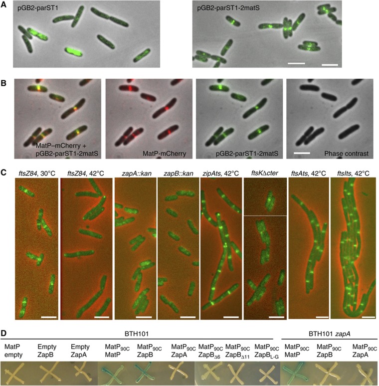
MatP interacts with ZapB. (A) Merged pictures of the pGBparST1 or the pGBparST1-2matS plasmid (green) and phase-contrast micrographs (grey) in MG1655 cells. (B) Montage of merged pictures of the MG1655matP–mCherry strain containing the pGBparST1-2matS plasmid. pGBparST1-2matS (green), MatP–mCherry (red) and phase-contrast microscopy (grey) are shown. (C) Localization of the pGBparST1-2matS plasmid (green) in MG1655ftsZ84, zapA, zapB, ftskΔC strains grown at 30°C and the MG1655ftsZ84, zipAts, ftsAts and ftsIts strains grown at 42°C for 1 h. Scale bars represent 3 μm. (D) Bacterial two-hybrid assay; the BTH101 and BTH101zapA strains containing the pKT25, pKT25MatP, pKT25ZapA, pKT25ZapB, pKT25ZapBΔ6, pKT25ZapBΔ11 or pKT25ZapBL-G and pUT18c or pUT18CMatP90C plasmids were grown in LB supplemented with kanamycine, ampicilin, IPTG and Xgal at 30°C; pictures were taken after 2 days of incubation at 30°C.

