Figure 2.
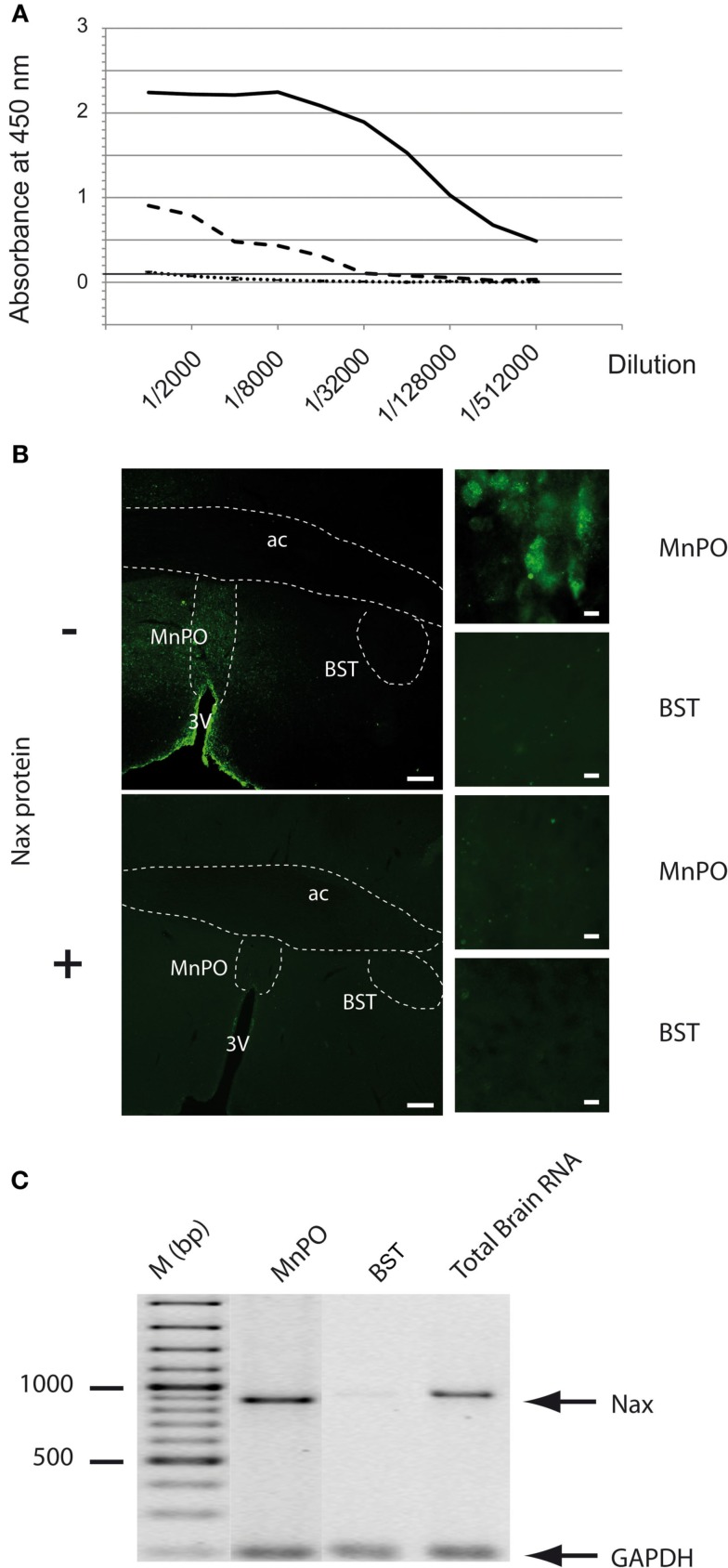
Specificity and validation of the anti-NaX antibody. (A) The immune serum collected from the rabbits was assessed by ELISA and revealed the presence of the NaX protein and not the GST tag. The microplate was coated with the NaX peptide (1 μg/well), incubated with pre-immune serum (dotted line), or double purified immune serum (black line: antibody titer >>1/512,000). To control purification efficiency, ELISA was also performed on GST protein with double purified immune serum (dashed line). The rabbit antibodies were revealed with goat anti-rabbit IgG, HRP conjugated secondary antibody and the absorbance was read at 450 nm. (B) The purified anti-NaX antibody (antibody concentration 1/250) was tested for immunohistochemical staining without (−) or with (+) preabsorption with the antigen peptide (50 μg/ml). Fluorescent immunostaining in the MnPO nucleus served as a positive control, whereas absence of immunostaining in the BST nucleus is a negative control. Scale bar: 200 μm (low magnification), 10 μm (high magnification). (C) The immunohistochemical staining was correlated with the presence of NaX mRNA revealed by RT-PCR experiment carried out on micropunched region of the rat MnPO and BST. Note that the expected size of amplified products was 883 bp for NaX and 200 bp for GAPDH (housekeeping gene). Commercial rat brain total RNA (Clontech Laboratories Inc.) was used as a positive control.

