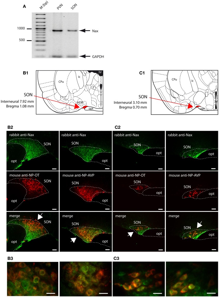
Figure 9.
NaX is expressed in the magnocellular neuroendocrine cells of the rat and mouse supraoptic nucleus. (A) NaX mRNA expression was revealed by RT-PCR experiment carried out on micropunched region of the SON and PVN. Note that the expected size of the amplified products was 883 bp for NaX and 200 bp for GAPDH (housekeeping gene). Schematic illustration of the SON in the rat (B1) and mouse (C1). The SON appears in red for better visualization. Representative distribution of fluorescent NaX immunostaining (green) in the magnocellular neurons of the SON obtained from the rat (B2) and mouse (C2). The neurochemical content of the cells expressing NaX was identified with the anti-oxytocin neurophysin and the anti-vasopressin neurophysin fluorescent immunostaining (red); scale bars: 50 μm. The arrow head points the top-right or the bottom-left corner of the inset, which represents a high magnification zone of the rat SON (B3) and the mouse SON (C3); scale bar: 20 μm. Not that NaX immunostaining was present in both vasopressinergic and oxytocinergic magnocellular cells in the rat and mouse.

