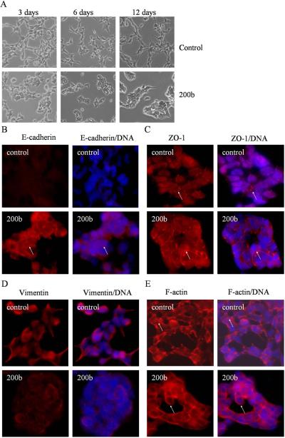
Fig. 4.
MiR-200b reverses EMT phenotype of PC3 PDGF-D cells. (A) Photographs of cells are shown: PC3 PDGF-D cells transfected with negative control miRNA exhibit a fibroblastic-type phenotype (upper panel), PC3 PDGF-D cells transfected with miR-200b display round-like epithelial cell shape and cells form a cluster (lower panel). Original magnification, 200 ×. After 21 days of transfection, PC3 PDGF-D cells transfected with negative control miRNA or miR-200b were immunostained for the expressions of E-cadherin (B), ZO-1 (C), vimentin (D), or stained with Alexa Fluor 594 phalloidin for F-actin (E) with DAPI for DNA to show cell nucleus, as described under method section. Arrows indicate changes in the expression or location of epithelial and mesenchymal markers. Original magnification, 200 ×.

