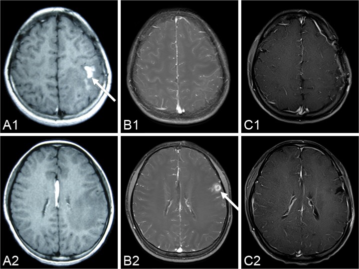
Figure 1.
A series of pre-operational MR images demonstrates the local migration of an irregular, enhanced lesion (arrow) in the left hemisphere (A1-A2) six months before the operation; (B1-B2) three days before the operation. The lesion disappears in the post-operation MR images over the two-year follow-up period (C1-C2) 26 months after the operation.

