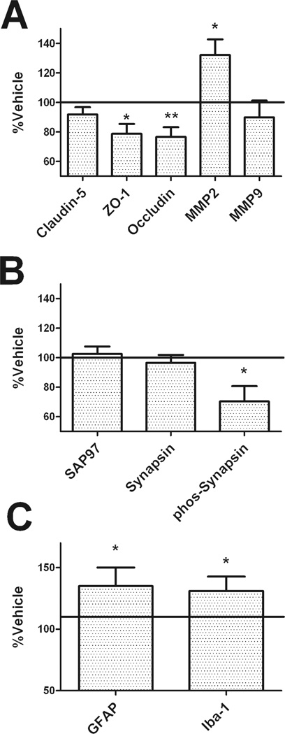
Figure 4. Lopinavir/ritonavir induces brain injury in mice.
Male C57BL/6 mice were treated daily with vehicle or lopinavir/ritonavir (150/37.5 mg/kg body weight) for 28 days, after which markers of cerebrovascular integrity, synaptic density, and reactive gliosis were evaluated in tissue homogenates prepared from the frontal cortex as described in Methods. Data depict mean ± SEM expression in lopinavir/ritonavir-treated mice presented as % vehicle (100% line) on graph. Data were obtained from 9–20 mice/group, and were analyzed by 2-tailed, unpaired t-tests. (A) Expression of the tight junction proteins claudin-5, ZO-1, and occludin; and the matrix metalloproteinases MMP2 and MMP9. * and ** indicate significant (p < 0.05 and 0.01, respectively) changes in expression in lopinavir/ritonavir-treated mice as compared to vehicle. (B) Expression of the post-synaptic marker synapse associated protein 97 (SAP97), the pre-synaptic protein synapsin 1, and phosphorylated synapsin 1. * indicates significant (p < 0.05) the significant decrease in phosphorylated synapsin 1 expression in lopinavir/ritonavir-treated mice. (C) Expression of the glial markers glial fibrillary acidic protein (GFAP) and Iba-1. * indicates significant (p < 0.05) increases in GFAP and Iba-1 expression in lopinavir/ritonavir-treated mice.

