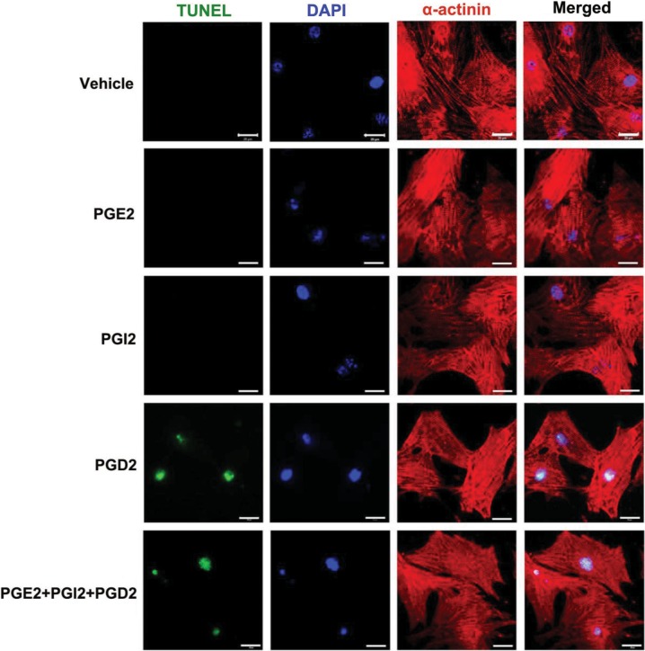
Figure 3.
Effects of prostanoids on cardiac myocyte apoptosis. Isolated neonatal cardiac myocytes were cultured for 48 h and serum starved for 24 h before starting the experiments. PGD2, PGI2, and PGE2 (Cayman Chemical) were dissolved in ethanol as 1000× stock solution and were added into culture medium at a concentration of 10 μM individually or in combination (10 μM each). In control experiments, cells were treated with the same volume of vehicle (ethanol) that was also diluted 1000× in the culture medium. After 6h of incubation, cells were fixed with 4% paraformaldehyde, treated with proteinase K, permeabilized in 0.01% Triton-X-100. Permeabilized cells and sections were then incubated with TUNEL reaction mixture (Roche In Situ Cell Death Detection Kit, Fluorescein) at 37°C for 1h. This was immediately followed by incubation with primary antibody for α-actinin (1:500, Sigma) overnight at 4°C, then Alex-Fluor-555-labeled secondary antibody (1:300, Invitrogen). The nuclei were counter stained by DAPI in the mounting media. Scale bar = 20 μm.

