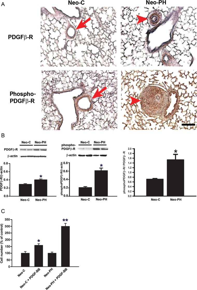
Figure 1.
Chronic hypoxia exposure stimulates an increase in the levels of PDGFβ-R, phosphoPDGFβ-R, and PDGF-BB-stimulated proliferative responses in PA adventitial fibroblasts. (A) Immunohistochemical staining of paraffin-embedded lung sections of Neo-C and Neo-PH calves was performed using antibodies against PDGFβ-R and phosphoPDGFβ-R. Arrows indicate PA adventitia compartments of Neo-C animals. Arrowheads indicate remodelled adventitial layers of PA of Neo-PH calves. Scale bar = 50 µm. Representative photomicrographs are presented. Staining was performed in lung sections of three different Neo-C and three different Neo-PH calves. (B) PDGFβ-R and phosphoPDGFβ-R levels were measured by western immunoblot analysis in growth-arrested PA adventitial fibroblasts isolated from Neo-C and Neo-PH calves. Densitometric quantification of the bands using NIH Image J program and normalized to β-actin is shown below each blot. pPDGFβ-R levels were also normalized to the level of PDGFβ-R in both Neo-C and Neo-PH cells. Values are mean ± SEM from three independent experiments where cells isolated from three different Neo-C and Neo-PH calves were used. *P< 0.01 vs. Neo-C value. (C) Quiescent fibroblasts were stimulated with PDGF-BB (25 ng/mL) for 48 h and then counted. The proliferation rate is represented as % of untreated cells. Data are mean ± SEM from three independent experiments using fibroblasts isolated from three different Neo-C and Neo-PH calves. *P< 0.01 vs. untreated Neo-C cells; **P< 0.01 vs. untreated Neo-PH cells and PDGF-BB-treated Neo-C cells.

