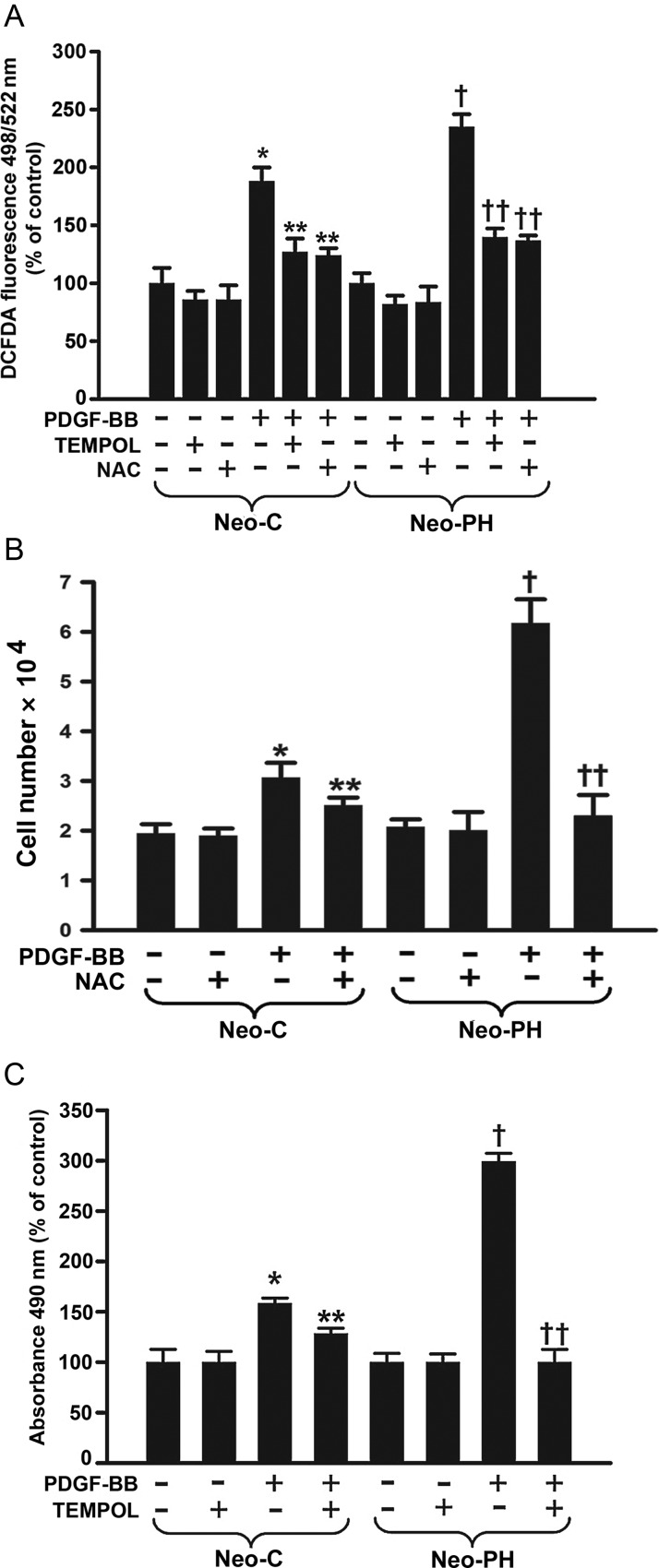
Figure 2.
PDGF-BB stimulates PA adventitial fibroblast proliferation through generation of intracellular ROS. (A) H2O2 levels were measured 2 h after PDGF-BB stimulation in both Neo-C and Neo-PH cells using DCFDA fluorescent dye and are represented as % of untreated cells. (B) Growth arrested cells were pretreated with N-acetyl cysteine (NAC) for 1 h, stimulated with PDGF-BB for 48h, and counted. (C) Quiescent fibroblasts were stimulated with PDGF-BB after pre-incubation with 4-Hydroxy-2,2,6,6-tetramethylpiperidinyloxy (TEMPOL), a SOD mimetic, for 1 h and proliferation assay was performed after 48 h. Data are mean ± SEM from three independent experiments using cells isolated from three different Neo-C and Neo-PH calves. *P< 0.001 vs. untreated Neo-C cells; **P< 0.01 vs. PDGF-BB treated Neo-C cells; †P< 0.01 vs. untreated Neo-PH cells; ††P< 0.01 vs. PDGF-BB treated Neo-PH cells.

