
Figure 6.
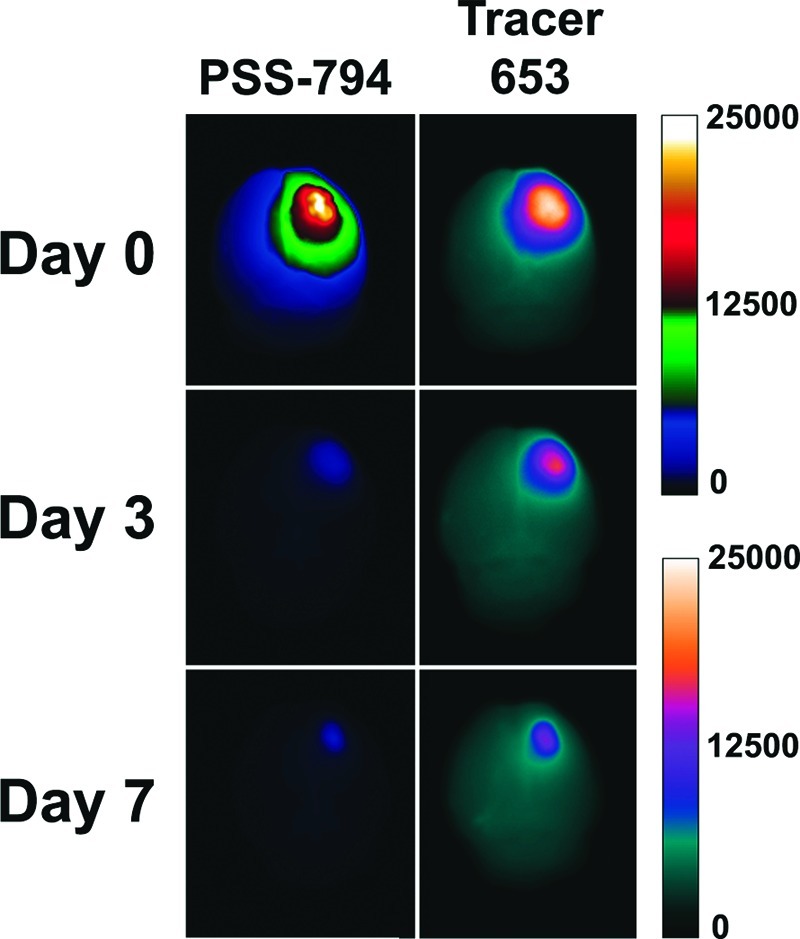
Multicolor fluorescence imaging of cell death and blood-brain-barrier disruption in cryoinjured brains. Three cohorts of hairless mice were given a 60 s brain cryoinjury. Mice were then injected with a single dose of PSS-794 either immediately following cryoinjury (Day 0), 3 days postcryoinjury (Day 3), or 7 days postcryoinjury (Day 7). Each mouse was also injected with Tracer-653 at 5 h post-PSS-794 injection. One hour after Tracer-653 injection, the mice were anesthetized and sacrificed. The brains were excised and placed in an epifluorescence imaging station for ex vivo imaging.