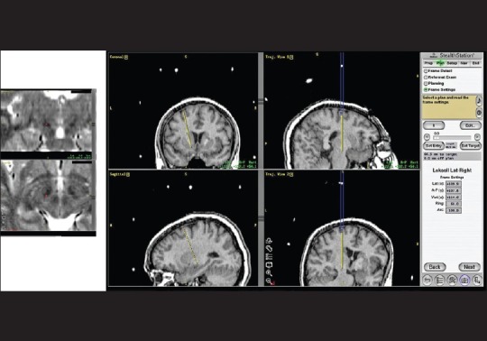
Figure 4.

In this example, the left subthalamic nucleus was targeted on T2 weighted stereotactic MRI (inset left). Stereotactic T1 volumetric images allowed selection of the entry point in the ipsilateral, pericoronal region right): Coronal and sagittal images are not sufficient to confirm avoidance of sulci and ventricles (left panel); this is best achieved by reformatting of the images in line with the planned trajectory (middle panel). The final target coordinates, together with the arc and ring angles then define the surgical trajectory (right). It should be noted that, prior to surgical planning, the fiducials were registered on the T2-weighted stereotactic MRI to avoid fusion errors during target selection. The entry point was selected on the basis of the volumetric T1 images fused to the T2 images and is therefore susceptible to errors of image fusion. However, inaccuracies are of less clinical relevance at the entry point than at the target point
