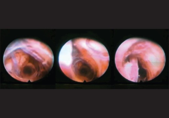
Figure 6.

Blunt probes tend to displace rather than rupture blood vessels crossing the surgical trajectory. These still shots are from an endoscopic video recorded during extraction of a small, 3 mm diameter blunt- tip endoscope from the brain. The left panel demonstrates the parenchyma and vessels in the distal brain track without evidence of haemorrhage. On the middle panel, a larger calibre vessel (arteriole) appears out of focus on the left side of the picture as the endoscope is withdrawn. The right panel confirms that the aforementioned vessel was in the path of the probe. The vessel had clearly been pushed aside by the advancing probe without damage or penetration causing haemorrhage (video courtesy of Prof Marwan Hariz)
