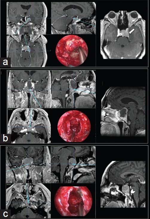
Figure 3.

Intraoperative neuronavigation images and each respective endoscopic endonasal intraoperative view. (a) Pituitary adenoma extending laterally and posteriorly to the left cavernous sinus and Meckel's cave – detail: T1-weighted contrast-enhanced axial MR image showing tumor invasion of the left cavernous sinus (arrow). (b) Large macroadenoma with a suprasellar extension and superior displacement of the optic chiasm. (c) Upper clivus invasion by a macroadenoma
