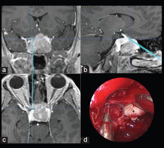
Figure 4.

Illustrative case: A 30-year-old male presented with headaches and progressive visual loss. Endocrine evaluation showed increased Thyrotropin (TSH) levels. MR imaging was performed and a macroadenoma, partially embracing the right carotid artery, was diagnosed. The neuronavigation system was used to assist the approach and to evaluate tumor proximity to the right carotid artery. Coronal (a), Sagittal (b) and Axial (c) intraoperative neuronavigation images of a transsphenoidal approach for a macroadenoma. (d) Endoscopic endonasal surgical view of left ICA localization during tumor (*) removal
