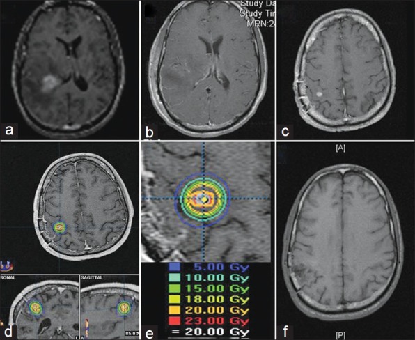
Figure 1.

The patient was 47 years old who presented with headache and dysphasia. Brain MRI before surgery (a) shows a periventricular contrast-enhancing mass with surrounding edema. Postoperative MRI (b) shows gross total resection and pathology confirmed glioblastoma. The patient underwent XRT and concomitant TMZ. Two months after adjuvant therapy, follow-up MRI (c) shows a small recurrent nodule outside the tumor cavity. This was targeted with SRS. Isodose lines around the lesion (d) treated with 20 Gy at the 85% isodose line (e). Follow-up MRI (f) shows radiographic control up to 19 months later
