Fig. 8.
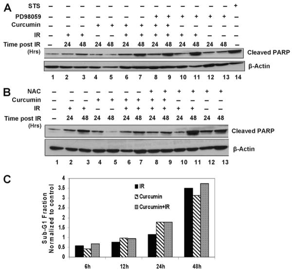
Curcumin induced apoptosis is through IR-induced ROS and prolonged ERK activation. A, HeLa cells were pretreated with DMSO, 10 μM curcumin (8 h), 5 mM NAC (6 h), or NAC treatment during the last 6 h of curcumin treatment. The cells were irradiated at 6 Gy and harvested 24 or 48 h after IR. Whole-cell lysates were analyzed by Western blot using antibodies against cleaved PARP and β-Actin. B, HeLa cells were pre-treated with DMSO, 10 μM curcumin (8 h), 50 μM PD98059 (2 h), or PD98059 treatment during the last 2 h of curcumin treatment. The cells were irradiated at 6 Gy and harvested 24 or 48 h after IR. Whole-cell lysates were analyzed by Western blot using antibodies against cleaved PARP and β-Actin. Lysate from cells treated with 1 μM staurosporine for 2 h was used a positive control for cleaved PARP. C, HeLa cells were treated with DMSO or curcumin followed by mock irradiation or 6-Gy dose of IR. The cells were fixed and treated with PI/RNase containing solution as described under Materials and Methods and subsequently analyzed by flow cytometry. Results represent the values of sub-G1 fractions normalized to un-irradiated controls.

