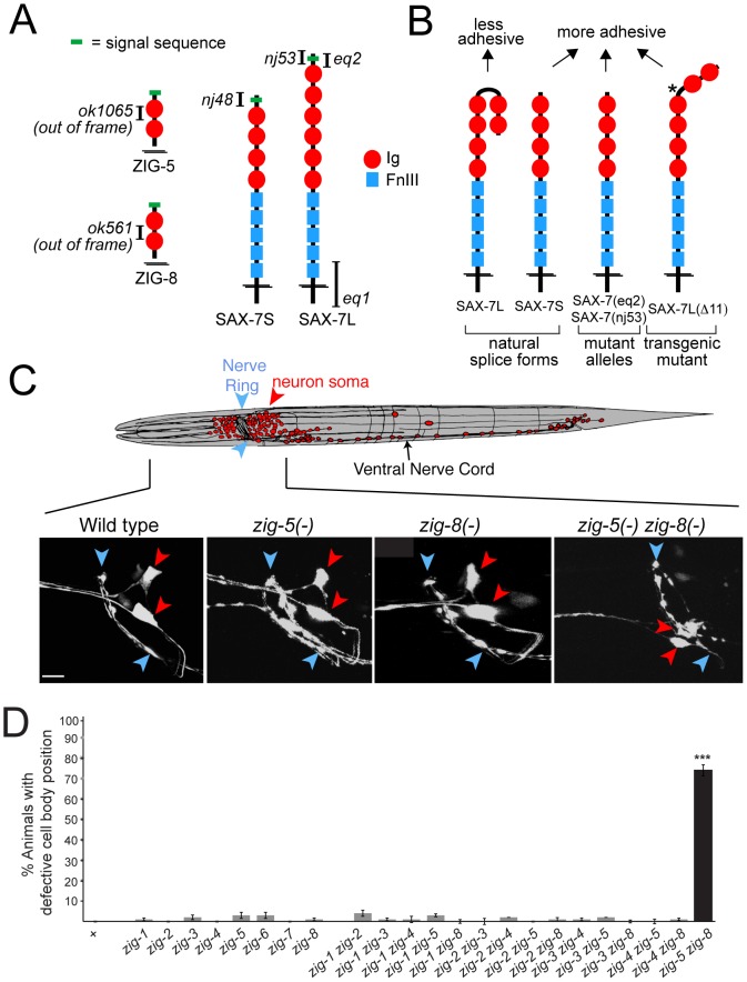
Figure 1. Neuronal maintenance factors and the defects caused by their removal.
(A) Schematic protein structures and alleles used in this study. (B) Summary of previous in vitro and in vivo adhesion studies [6], [7]. Star indicates a shortened hinge region which prevents formation of the horseshoe configuration [7]. (C) ASI and ASH neuronal displacements observed in zig-5(ok1065) and zig-8(ok561) single and double mutant adult animals with the oyIs14 reporter transgene. Blue arrowheads indicate position of the nerve ring and red arrowheads position of neuronal soma, which is scored relative to position of the nerve ring (wild type: behind nerve ring; mutant: on top of to nerve ring). Anterior to left, dorsal on top. Scale bar is 5 µm. (D) Quantification of ASI and ASH neuronal displacement in single and double mutants of the zig gene family. Alleles are described in [11]. Error bars indicate s.e.p.. Proportions of different animal populations were compared using the z-test. “*” indicates p<0.001.

