
Figure 8. Lamellocyte WGA staining was reduced in L. victoriae attacked larvae and restored by Mgat1 overexpression.
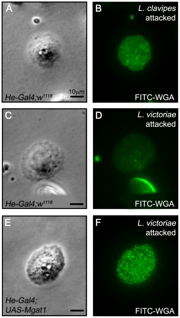
Paired brightfield and FITC images of hemocytes stained with FITC-WGA 24–48 hours following attack by the indicated wasp. (A–D) Lamellocytes from He-Gal4;w1118 larvae were WGA+ following L. clavipes attack (A,B), but were WGA− following L. victoriae attack (C,D). (E,F) The WGA+ staining was restored to L. victoriae attacked larvae by hemocyte specific expression of Mgat1 in He-Gal4;UAS-Mgat1 larvae. Scale bars indicate 10 µm.