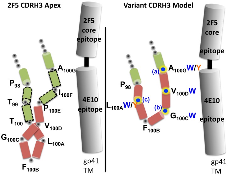
Figure 4. Schematic representation of the 2F5 wt and loop-shortened CDRH3 and approximate location of the MPER.
Left, the wt 2F5 CDRH3 apex is shown as a schematic representation based upon the mAb structure. Residues P98 to A100G are shown with each residue represented by a colored cylinder. On the right, a schematic of the length-altered version, where residues T99,T100, P100E and I100F , represented by the green cylinders with the discontinuous border, were deleted. The red residues, L100A, F100B, G100C and V100D, were maintained for the length alterations as described in the text. The W and Y substitutions are noted next to the original residue in the variant schematic depiction (Rosetta modeling of a variant CDRH3 was performed, but clashes with peptide binding were assessed to not be compatible with the predicted conformations). The letters a, b and c represent the locations of the second W substitutions to generate antibody variants r4W2a, b and c, respectively.

