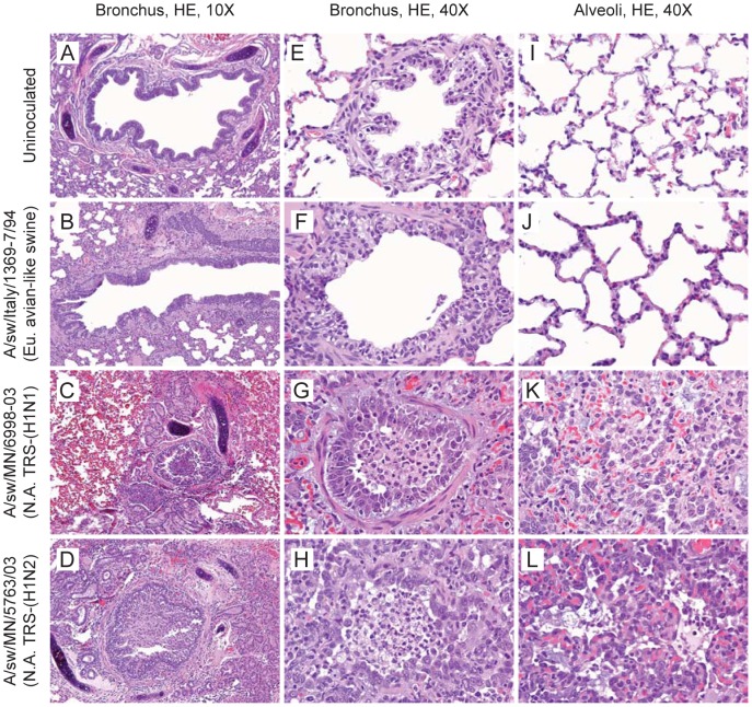
Figure 2. Histopathology of ferret lung tissue.
Lung tissue of control (un-inoculated) and virus-inoculated ferrets was collected on day 5 p.i. Formalin-fixed, paraffin-embedded 5-µm sections were stained with hematoxylin and eosin and microscopically examined in a blinded fashion. Representative images show bronchi (A–D), bronchioles (E–H), and alveoli (I–L) from un-inoculated (A,E,I) and virus-inoculated ferrets. The two North American TRS viruses (C,G,K and D,H,L) caused bronchitis, bronchiolitis, alveolitis, and alveolar wall interstitial changes. The bronchitis featured intraluminal granulocytes and/or mucus, bronchial epithelial hyperplasia with submucosal mucus gland loss, and mixed inflammatory-cell infiltrates. The bronchiolitis featured intraluminal cellular debris, sloughed epithelial cells, and inflammatory cells (macrophages and/or granulocytes). The peribronchiolar alveoli contained mixed inflammatory cell infiltrates and foci of pneumocyte hyperplasia. The Eurasian avian-like swine virus (B,F,J) caused morphologic changes similar to those caused by the TRS viruses but far less severe.

