
Figure 1. PDB structures.
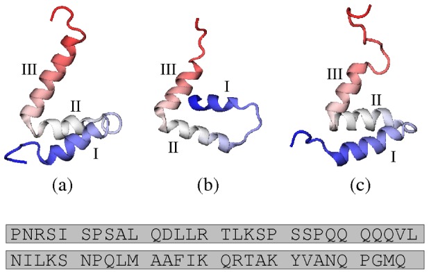
(a) Model 1 from 1KBH, the structure of NCBD in complex with ACTR [40]; (b) Chain C from 1ZOQ, the structure of NCBD in complex with IRF-3 [43] (the asymmetric unit comprises two NCBD molecules and two IRF-3 molecules); (c) Model 1 from 2KKJ, the core unbound conformer of NCBD [46]. In this work, we choose these conformations to represent the three PDB structures. Helical regions I, II and III are indicated for each structure. Beneath is the amino acid sequence of NCBD. We follow reference [46] in numbering the residues of NCBD sequentially from 1 to 59. This corresponds to residues 2059–2117 of mouse CBP in 2KKJ and 1KBH (also numbered 48–106 in 1KBH). Structure 1ZOQ contains residues 2065–2111 of human CBP, whose sequence matches residues 2066–2112 of 2KKJ and 1KBH; therefore, 1ZOQ contains residues 8–54 according to the numbering system that we use.