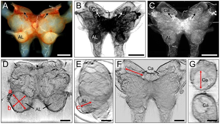
Figure 2. Evaluation of size changes of the antennal lobe and mushroom body with Scanning Laser Optical Tomography (SLOTy).
A. Frontal view of the untreated locust brain (exclusive optic lobes) under reflected light. The mushroom body calyces are hardly visible (arrows). B,C. Raw data of 3D transmission-(B) and scattered light (C) projections of the locust brain. Brains were cleared with glycerol. Note that mushroom body calyces are clearly visible (arrows in B,C). D–G. Reconstructed 2D optical sections of transmission projections. D. Frontal section depicting left (untreated side) and right (ablated side) antennal lobe 7 days after ablation of the right antenna. Note that the right (deafferented) antennal lobe is smaller than the left (untreated) antennal lobe. E. Side view of brain with left antennal lobe. Antennal lobe size was approximated as an ellipsoidal volume V = π/6abc (D,E). F,G. Frontal section (F) and horizontal section (G) depicting mushroom body calyces. Calyx width was measured in frontal sections (red line in F). AL, antennal lobe; Ca, calyx. Scale bars = 400 µm in A–C; 200 µm in D–G.

