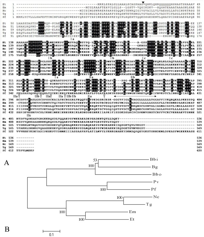
Figure 1. Characterization of the EtAMA1 gene.
(A) Multiple-sequence alignment of AMA1 proteins from E. tenella (Et), E. maxima (Em), N. caninum (Nc), T. gondii (Tg) and P. falciparum (Pf). Strictly conserved residues are indicated with a black background, and five different cysteine residues with species of Plasmodium in DIII are indicated with a gray arrowhead. The cleavage site of the EtAMA1 putative signal peptide is indicated by an arrowhead, and the transmembrane region is indicated by ( ); cysteine residues that formed disulfide bonds in EtAMA1 are indicated by domain (I, II and III) and bond (a, b and c) designations according to TgAMA1. (B) Phylogenetic tree of AMA1 proteins from E. maxima (Em), E. tenella (Et), N. caninum (Nc), T. gondii (Tg), P. falciparum (Pf), P. vivax (Pv), B. bovis (Bbo), B. gibsoni (Bg),and P. bigemina (Pbi).

