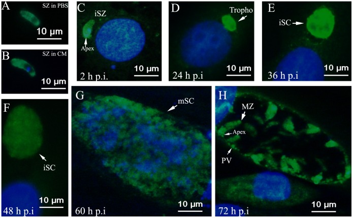
Figure 5. EtAMA1 localization in DF-1 cell infection as visualized by immunofluorescence analysis.
Details of parasites immuno-stained with anti-rEtAMA1 antibodies, visualized with FITC (green) and counter-stained with DAPI (blue). (A) Sporozoites (SZ) were incubated in PBS or (B) complete medium (CM) at 41°C. Infected DF-1 cells were collected at different time points p.i. (C) 2-h p.i., intracellular sporozoites (iSZ); (D) 24-h p.i., intracellular trophozoite (Tropho); (E) 36-h p. i., immature schizont (iSc); (F) 48-h p.i., immature schizont (iSc); (G) 60-h p.i., mature schizont (mSc); (H) 72-h p.i., merozoites (MZ).

