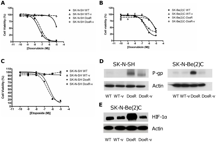
Figure 1. Drug resistance in the SK-N-SH and SK-N-Be(2)C cells.
Doxorubicin resistance was assessed by MTT cell proliferation assay in the (a) SK-N-SH and (b) SK-N-Be(2)C cell lines. WT and WT-v proliferation was reduced by increasing concentrations of doxorubicin whilst the DoxR and DoxR-v cells were equally resistant to doxorubicin therapy. (c) Cross resistance to etoposide was likewise confirmed in the SK-N-SH cell line. (d) Western immunoblotting demonstrated significantly greater upregulation of P-glycoprotein (P-gp) in the DoxR than the DoxR-v cells. (e) Likewise in the SK-N-Be(2)C cell line, western blot revealed a pattern of upregulation in hypoxia inducible factor -1α (HIF-1α) similar to that observed with P-gp.

