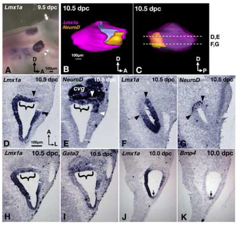
Fig. 3. Lmx1a expression influences neural subtype specification.

(A) Lmx1a expression in a whole-mount embryo. Lmx1a is not expressed in the antero-ventral region of the otocyst (arrowheads). (B) Lateral and (C) medial views of a 3-D reconstructed inner ear showing the relationships of Lmx1a (pink) and NeuroD (yellow) expression domains. The pink in (C) is rendered transparent to reveal the yellow area underneath. Asterisk indicates the neurogenic region that is devoid of Lmx1a expression. (D-G) Representative sections from ear shown in (B) and (C). Brackets indicate the neurogenic regions that do express Lmx1a. Arrowheads mark the neurogenic regions that do not express Lmx1a. (H,I) adjacent sections probed for Lmx1a (H) and Gata3 (I). (J,K) adjacent sections probed for Lmx1a (J), and Bmp4 (K), showing Lmx1a expression in the Bmp4-positive posterior crista (arrows). Sections are rotated 90° clockwise from orientation in (C). Orientation and scale bar in D apply to E–K.
