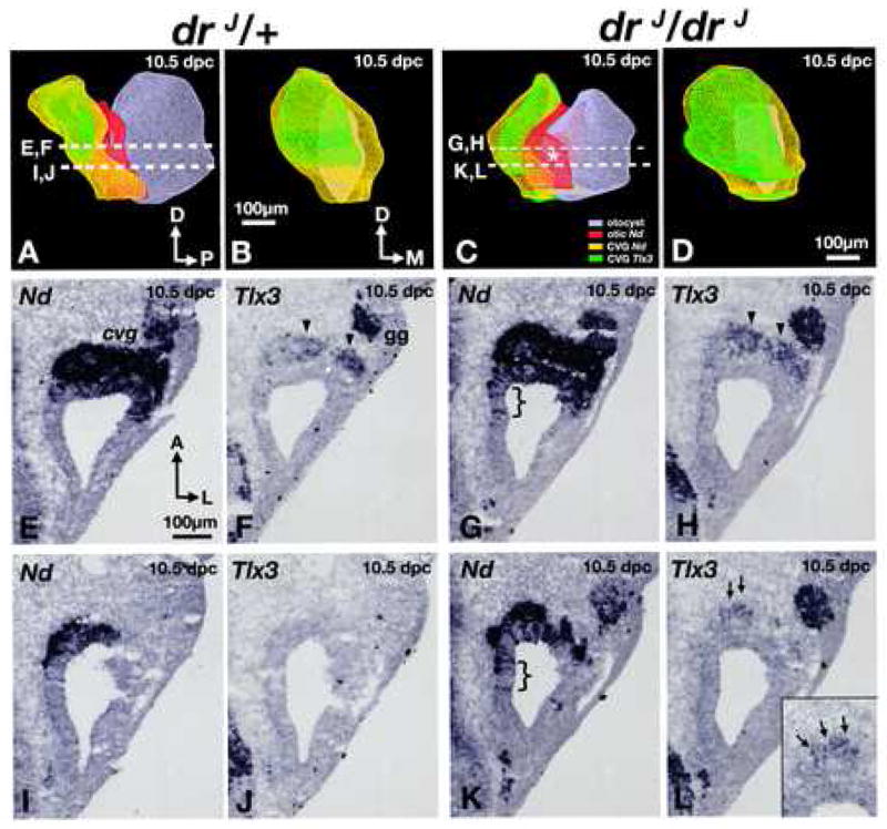
Fig. 4. Ganglion malformations in drJ/drJ mutants.

(A,C) Antero-medial views of 3-D reconstructed, right inner ears (grey) of (A) heterozygous and (C) homozygous drJ/drJ mutants. NeuroD expression within the otocyst is red, and expression of NeuroD and Tlx3 in the CVG are yellow and green, respectively. Asterisk represents a medial expansion of the NeuroD domain in the dreher otocyst. (B,D) Anterior views of the CVG in (A) and (C), respectively. (E,F) and (I,J) are representative sections from the drJ/+ specimen shown in (A). Comparable sections from the drJ/drJ specimen in (C) are shown in (G,H) and (K,L). (E-H) At this level, there is an expansion of the neurogenic domain (G; bracket) and more Tlx3 expression in the CVG of drJ/drJ compared to drJ/+ (H; arrowheads). (I-J) Slightly ventral, where there is only residual neurogenic domain that is Tlx3 negative in drJ/+, there is a broader neurogenic domain in drJ/drJ mutants (K; bracket) and some Tlx3-positive neuroblasts can be detected (L; arrows; inset). Sections are rotated 90° clockwise from orientation in (A or C). gg, geniculate ganglion. Orientation and scale bar in E apply to F-L.
