
Fig. 7. Abnormal gene expression patterns in the cochlear duct and cristae of drJ/drJ mutants.
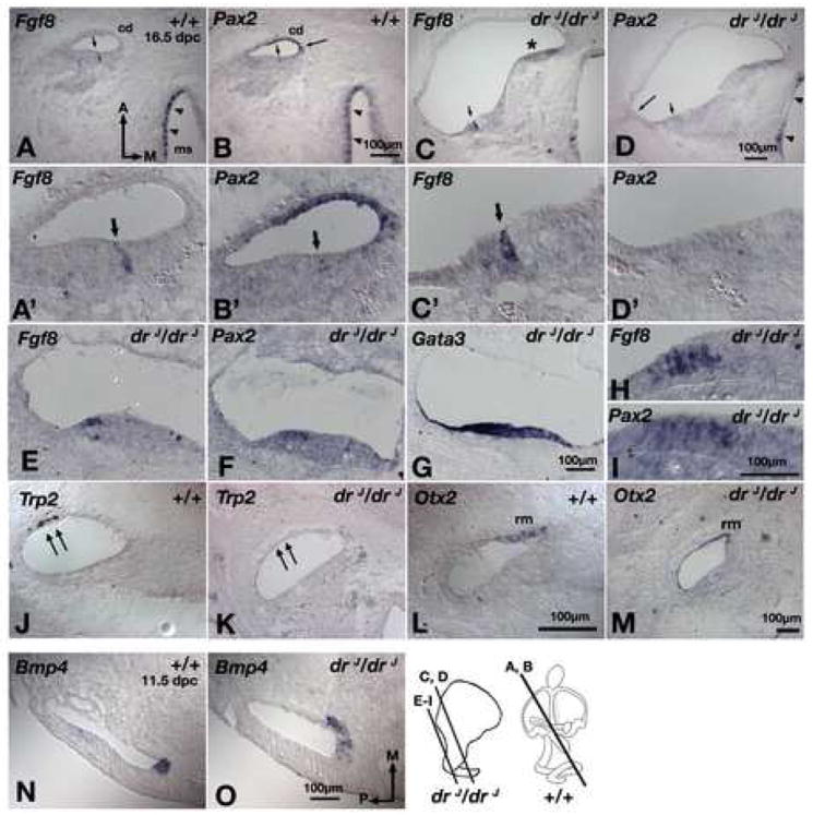
(A,B) Adjacent sections of wildtype inner ears showing Fgf8 (A) is expressed in the inner hair cells of the organ of Corti (arrow) and hair cells of the macula of the saccule (ms; arrowheads). Pax2 (B) is expressed in the stria vascularis (long arrow), hair cells of the vestibule (arrowheads), but expression in the hair cells of the organ of Corti is barely detectable (short arrow). (C,D) Adjacent sections of the apical region of drJ/drJ cochlear duct showing Fgf8 (C) is expressed in the inner hair cells of the organ of Corti, but Pax2 (D) is in neither the stria vascularis (long arrow) nor sensory hair cells (short arrow). Asterisk indicates an area where the tissue is folded. (A’-D’) Higher magnification of sections shown in (A-D). (E,F,G) Adjacent sections of the basal region of the drJ/drJ cochlear duct showing high numbers of hair cells expressing Fgf8 (E) and Pax2 (F) in a Gata3-positive, cochlear region (G). (H,I) Higher magnification of sections shown in (E) and (F), respectively. (J,L) In wildtype cochlear duct, Trp2 (J) and Otx2 (L) are expressed in the stria vascularis (double arrows) and Reissner’s membrane (rm), respectively. In drJ/drJ cochlear duct, Pax2 (D, long arrow) and Trp2 (K) in the stria vascularis are not detected, but Otx2 expression in the Reissner’s membrane is evident (M). (N,O) Bmp4 expression in the presumptive anterior crista is expanded medially in drJ/drJ inner ears (O) compared to wildtype (N). cd, cochlear duct. Scale bars: (A,B);(C,D);(E-G);(H,I);(J-L);(N,O)