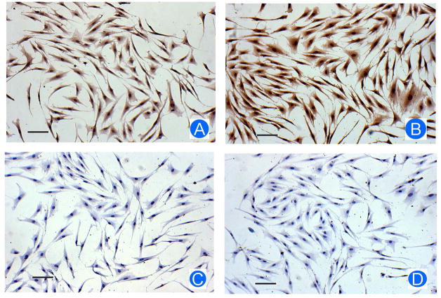
Figure 2.
Reduction of SFRP-1 protein in the dental follicle cells by CSF-1. After being treated with CSF-1, the dental follicle cells were immunostained for SFRP-1 protein level. The treated cells showed a reduced staining of SFRP-1 protein (A) as compared with control cells without treatment (B). In the staining controls in which primary antibody was replaced by IgG, no staining was observed in the cells treated with CSF-1 (C) or the cells without treatment (D). Scale bar: 50 μm.

