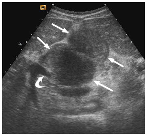
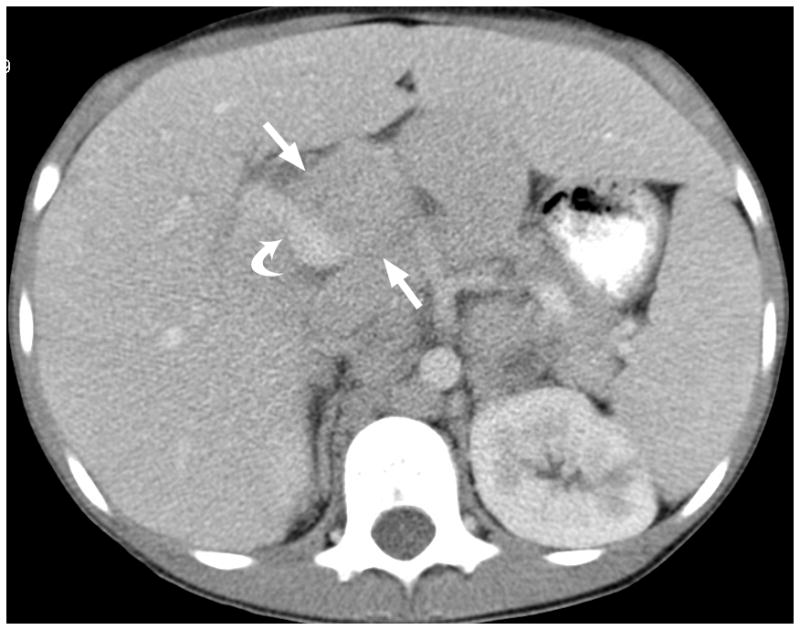
Fig. 2.


12-year-old boy with hepatocellular carcinoma and local nodal metastasis. (a) Non–contrast-enhanced transverse US image near the porta hepatis shows enlarged metastatic nodes (arrows) and main portal vein (curved arrow). (b) After administration of 0.56 mL of perflutren contrast agent, the main portal vein is opacified (curved arrow) and the relationship between the portal vein and adjacent node (straight arrows) is well defined. (c) Contrast-enhanced CT image at the level of the porta hepatis shows the enlarged node (arrows) adjacent to the main portal vein (curved arrow) as demonstrated by CEUS
