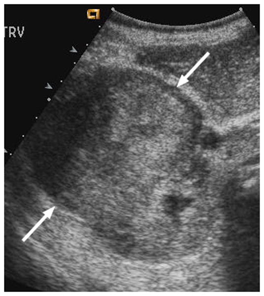
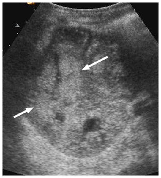
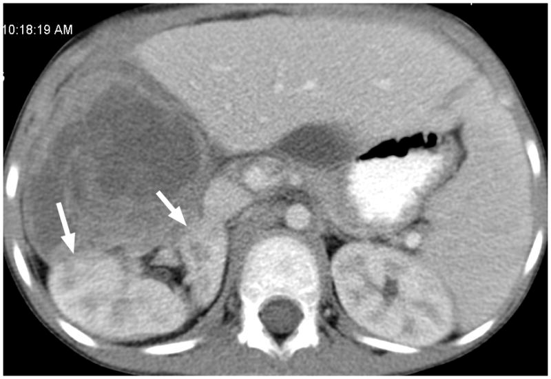
Fig. 3.


6-year-old boy with Wilms tumor. (a) Non–contrast-enhanced transverse US image of the primary right renal tumor (arrows). (b) After administration of 0.25 mL of perflutren contrast agent, there is slight enhancement in the lateral aspect of the tumor (arrows). Note that tumor margin is not well defined on pre- or post-contrast US images. (c) Contrast-enhanced CT image of the tumor better defines the interface between tumor and normal renal parenchyma (arrows)
