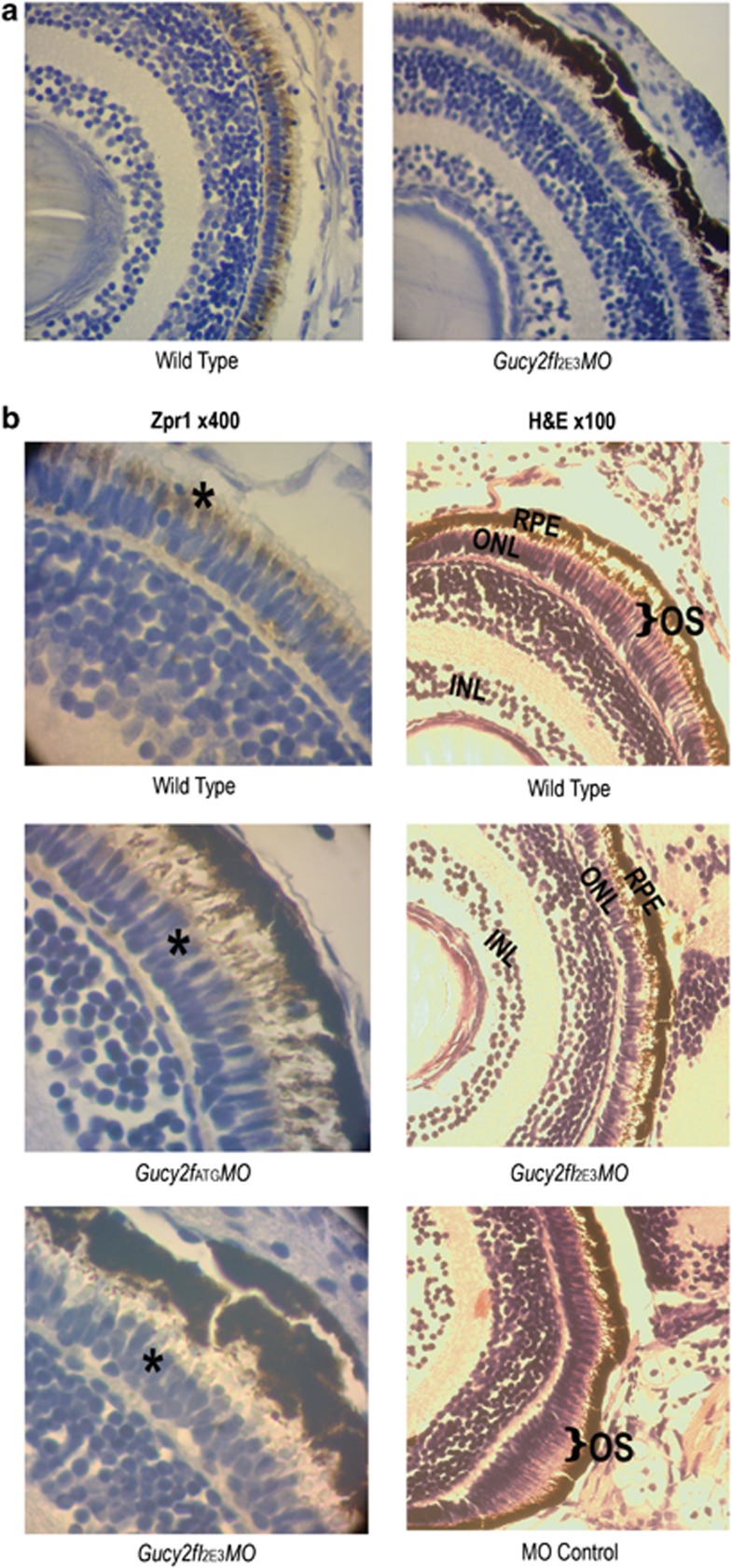
Figure 4.
Histological findings reveal that Gucy2f knockdown leads to retinal dystrophy. (a) First row: overall view of the retina demonstrates abundant zpr1 staining in wild-type larvae, absent in Gucy2fI2E3MO. (b) Left column: representative sections ( × 400) following zpr-1 antibody staining of double cones reveals (brown) double-cone staining (asterix) in the wild type in contrast to reduced staining in Gucy2fATGMO and in Gucy2fI2E3MO larvae. Right column: light microscopy, hematoxylin–eosin stain (H&E, × 100. Wild-type zebrafish larvae reveal normal retinal structures and development of the photoreceptor layers. Black bracket indicates length of photoreceptor outer segments. Gucy2fI2E3MO larvae display shortening of photoreceptor outer segments (no bracket). In the MO control, note the normal outer segments (black brackets).

