Fig. 7.
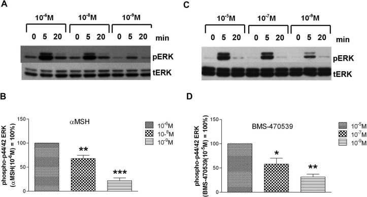
Cells expressing the wild-type MC1R show concentration-dependent transient increases in MAPK activation in response to αMSH and BMS-470539. HEK293 cells were transfected with cDNA encoding the wild-type MC1R. Forty-eight hours after transfection, the cells were stimulated with the indicated concentrations of either αMSH or BMS-470539 for 5 or 20 min, or cells were not treated with ligand (0 min). After stimulation, cells were lysed and levels of phospho-ERK (pERK) and total ERK (tERK) measured by Western blot analysis as described under Materials and Methods. A, a representative Western blot illustrates the concentration- and time-dependent effects of αMSH on phospho-ERK levels. B, quantitation of the Western blot signal 5 min poststimulation. Data are expressed as a percentage of the effect induced by 10−6 M αMSH. Comparisons were made relative to this maximal value: **, p < 0.01; ***, p < 0.001. Each bar represents the mean ± S.E.M from at least four independent experiments. C, a representative Western blot illustrates the concentration- and time-dependent effects of BMS-470539 on phospho-ERK levels. D, quantitation of the Western blot signal 5 min poststimulation. Data are expressed as a percentage of the effect induced by 10−5 M BMS-470539. Comparisons were made relative to this maximal value: *, p < 0.05; **, p < 0.01. Each bar represents the mean ± S.E.M from at least four independent experiments.

