
Fig. 2.
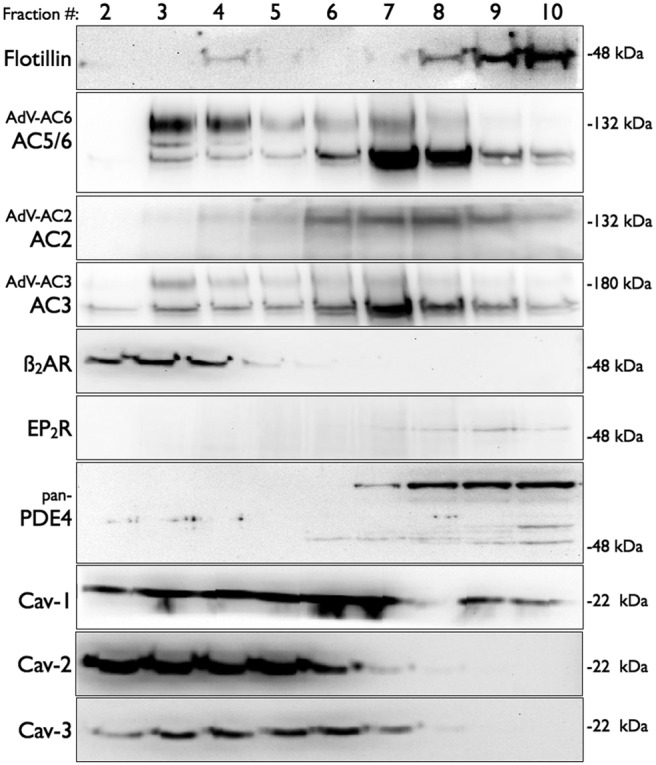
Immunoblot analysis of fractions from lipid raft isolation from mBSMCs. Cells were fractionated by using a nondetergent method and separated by sucrose density centrifugation (see Materials and Methods). Gradients were collected in 10 0.5-ml fractions and analyzed for appropriate separation of marker proteins (data not shown). Fractions were separated by SDS-polyacrylamide gel electrophoresis and analyzed by immunoblotting by using the indicated primary antibody. In some studies, cells were incubated with recombinant adenoviruses expressing AC2, AC3, or AC6 for 24 h. Shown are regions of the gels at the approximate molecular mass of the expected immunoreactive band. Images shown are representative of three to five experiments. Cav, caveolin.