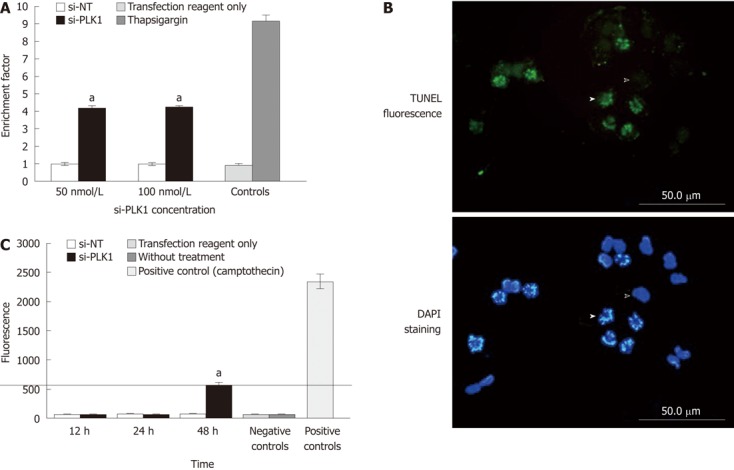
Figure 3.

Increased apoptosis, apoptosis by terminal deoxynucleotidyl transferase dUTP nick end labeling staining and caspase 3 activity after knockdown of polo-like kinase 1. A: Increased apoptosis; apoptosis measured by enrichment factor [enrichment factor (sample absorbance/control absorbance) > 1 indicates nuclear fragmentation]. Huh-7 cells transfected with short-interfering polo-like kinase 1 (si-PLK1) showed a 4-fold increase in enrichment factor compared with short-interfering non-targeting (si-NT) or the negative control, no difference was seen between the 50 nmol/L and 100 nmol/L si-PLK1. Thapsigargin (5 μmol/L) treated Huh-7 cells were positive controls. Data are shown as mean ± SE, using the Student t-test (aP < 0.05); B: Terminal deoxynucleotidyl transferase dUTP nick end labeling (TUNEL) fluorescence images showing fragmented DNA labeled with fluorescein-12-dUTP using recombinant terminal deoxynucleotidyl transferase (upper image). The lower image is the corresponding 4’,6-diamidino-2-phenylindole (DAPI) stain showing an apoptotic Huh-7 cell (solid arrowhead) and a non-apoptotic Huh-7 cell (hollow arrowhead). The TUNEL staining colocalized the fragmented chromosomes to apoptotic cells while is absent in non-apoptotic cells with intact chromosomes. Huh-7 cells were processed for fluorescence imaging after 24 h of transfection with 50 nmol/L si-PLK1; C: Caspase 3 activity was measured using a fluorometric immunosorbent enzyme assay from Huh-7 cells transfected with 50 nmol/L si-PLK1 showed no change after 12 and 24 h, with an increase to just over 500 U at 48 h compared with si-NT and negative controls. Positive controls showed values over 2350 U (cell lysates from U937 cells treated with camptothecin, supplied with the kit). Data are shown as mean ± SE, using the Student t-test (aP < 0.05).
