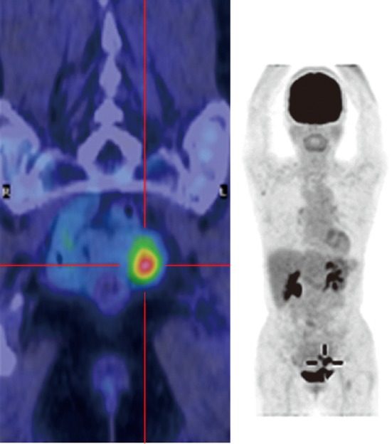
Figure 2.

Fluorodeoxyglucose-positron emission tomography computed tomography imaging. A fluorodeoxyglucose-positron emission tomography computed tomography scan showing increased fluorodeoxyglucose (FDG) uptake in the affected portion of the sigmoid colon (maximum standardized uptake value: 5.6 in the early phase and 7.4 in the late phase), but no increase in FDG uptake was observed in the surrounding lymph nodes or distant organs.
