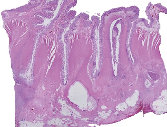
Figure 6.

Pathological examination of the resected specimen. Pathological examination of the resected specimen revealed no change in the mucosal epithelial surface, but multiple diverticula penetrated the muscularis propria with a marked thickening of the muscularis propria, fibrous thickening of interstitial tissue, severe infiltration of inflammatory cells, and a cystic change that contained inflammatory exudate and abscess formation.
