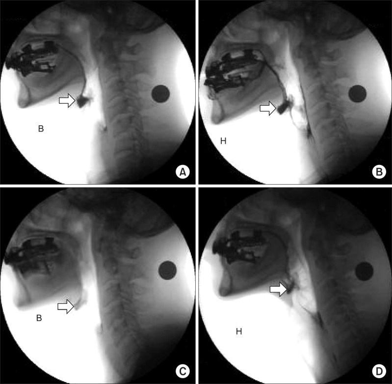
Fig. 2.
Videofluoroscopic swallowing study images were taken after swallowing bananas (A, C) and cookies (B, D). (A, B) the initial video fluoroscopic swallowing study demonstrates a moderate amount of residue in the valleculae (arrow). After 10 sessions of neuro muscular electrical stimulation, the follow-up VFSS was performed. (C, D) each examination shows a lesser amount of residue in the valleculae (arrow) compared with that in the previous test (B: banana, H: cookies).

