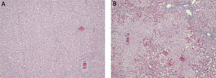
Figure 3.
Histopathological picture of the liver in SDR. A – Lack of morphological changes in the liver from sham SDR rats (group 4) at 48 h after saline injection. Haematoxilin and eosin staining, light microscope, magnification 10×. B – Massive necrosis of hepatocytes in the liver from tested SDR rats (group 5) at 48 h after galactosamine injection. Haematoxilin and eosin staining, light microscope, magnification 10×

