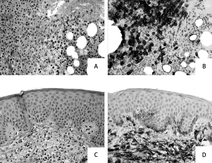
Figure 5.
Histopathological examination of the bone marrow and the skin samples. Biopsies from a patient with systemic mastocytosis. A, B – Bone marrow biopsy reveals focal mast cell accumulation. This fulfils the criterion of systemic mastocytosis (more than 15 mast cells per aggregate). Haematoxylin and eosin (A) and CD117 staining (B). C, D – Perivascular mast cell infiltrates in the upper portion of the dermis. Haematoxylin and eosin (C) and CD117 staining (D)

