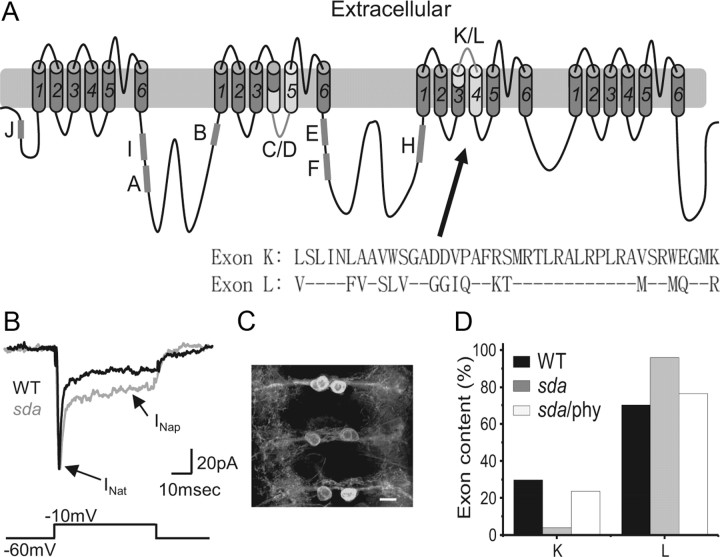
Figure 1.
Splicing of alternate exons K and L is altered in the sda mutant. A, Schematic of the DmNav transcript highlighting common spliced exons. Exons J, I, A, B, E, F, and H are subject to cassette-based splicing (i.e., can be present or absent), whereas exons C/D and K/L are mutually exclusive (i.e., one or other are present but not both). Inset, Exons K and L differ by 16 aa. B, Whole-cell voltage-clamp recordings from a third-instar aCC neuron reveals an increased INap in sda compared with WT. In contrast, INat is not different (for a full description, see Marley and Baines, 2011). C, GAL4RRa is sufficient to express GFP (UAS–GFPCD8 shown) in only aCC neurons by late-stage, wall-climbing third-instar larvae. A wide-field deconvolved fluorescent image shows three segments of the ventral nerve cord each containing two aCC motoneurons. Anterior is to the top. Scale bar, 10 μm. D, Analysis of splicing of DmNav, in aCC neurons isolated by FACS, shows that inclusion of exon L is greatly increased in the sda mutant (70.2 vs 96%, p ≤ 0.01). Preexposure of sda larvae to the antiepileptic drug phenytoin (phy) is sufficient to partially rescue this change (76.5%, p ≤ 0.05). Only functional DmNav splice variants are included in this analysis. Thus, DmNav69, in which K and L are coexpressed in the sda mutant, is not included because it is nonfunctional (see Materials and Methods).

