Figure 7.
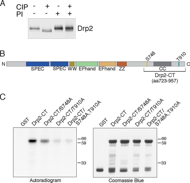
Drp2 is phosphorylated. A, Western blots of nerve lysates treated with calf intestinal phosphatase (CIP) with or without phosphatase inhibitors (PI) shows that Drp2 is phosphorylated. B, Phosphopeptide mapping of Drp2 by mass spectrometry identified S748 and T910 as the primary sites of phosphorylation. C, In vitro phosphorylation of GST fusion proteins containing the C-terminal region of Drp2 (amino acids 723–957) with peripheral nerve lysates in the presence of [γ-32P]ATP. Mutation of either S748 or T910 to alanine reduced phosphorylation. Coomassie Blue staining shows the amounts of the GST proteins were comparable.

