Figure 2.
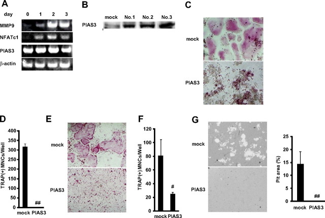
Overexpression of PIAS3 inhibits RANKL-mediated osteoclastogenesis in vitro. (A) Expression of MMP9, NFATc1, PIAS3, and β-actin mRNA. Total RNA from BMMs treated with 30 ng/mL M-CSF and 50 ng/mL RANKL for 0, 1, 2, and 3 days were measured by RT-PCR. (B) Western blot analysis of PIAS3 in RAW264.7 cells transduced with mock or PIAS3. (C) Analysis of osteoclast differentiation by TRAP staining. Mock- and PIAS3-transduced RAW264.7 cells were cultured for 3 days in the presence of 50 ng/mL RANKL. Cultured cells were fixed and stained for TRAP. (D) TRAP-positive MNCs having more than 3 nuclei were counted as osteoclast. (E) Analysis of osteoclast differentiation by TRAP staining. Mock- and PIAS3-transduced BMMs were cultured for 5 days in the presence of 30 ng/mL M-CSF and 50 ng/mL RANKL. Cultured cells were fixed and stained for TRAP. (F) TRAP-positive MNCs having more than 3 nuclei were counted as osteoclast. (G) Bone resorption assay using osteologic discs. Mock- or PIAS3-transduced RAW264.7 cells were cultured with 50 ng/mL RANKL on osteologic discs for 5 days. Pit area was quantified using ImageJ software. Error bars in all panels represent SD. #P < .05; ##P < .001.

