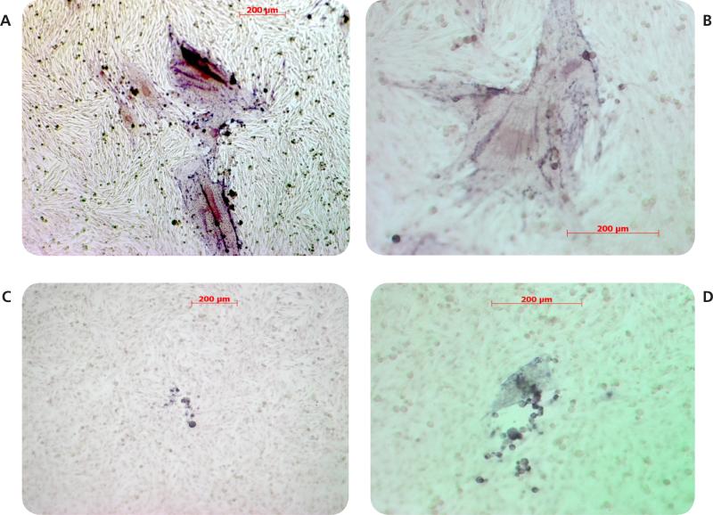
Figure 3.
Effect of honey on VZV plaque morphology in MeWo cells. MeWo cells were infected with VZV and maintained in 0% (panels A and B) or 6% (panels C and D) honey. Virus plaques were visualized by staining for VZV IE63 protein and photographed at 10x (panels A and C) or 20x (panels B and D) magnification.

