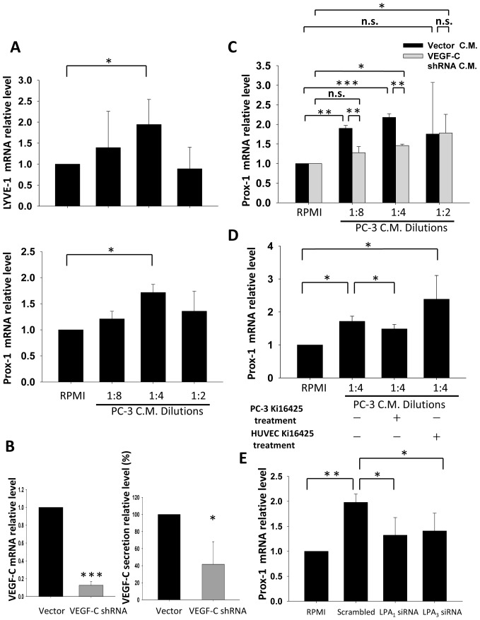
Figure 6. The role of LPA1 and LPA3 in PC-3 conditioned medium (CM) induced lymphatic marker expression.
(A) PC-3 cells were cultured in RPMI-only medium for 24 h after 16 h of starvation. The 24 h CM was collected and diluted to treat HUVECs for 4 h. After treatment, HUVEC lymphatic marker expression was observed. (B) PC-3 cells stably transfected with VEGF-C shRNA were tested for VEGF-C mRNA and secretion expression. (C) VEGF-C knockdown PC-3 CM was used to treat HUVECs following the procedure described above. (D) PC-3 cells were treated with 10 µM Ki16425 while being cultured in RPMI-only medium for 24 h before CM was used to treat HUVECs. Ki16425 was also used to test whether LPA existing in PC-3 CM is crucial for HUVEC lymphatic marker regulation. Ki16425 (10 µM) was pretreated for 1 h before PC-3 CM treatment of HUVECs. (E) CM from PC-3 cells transfected with LPA1 or LPA3 siRNA was used to treat HUVEC cells as procedures described above. Sample mRNA was reverse-transcribed and analyzed by a real-time PCR. Relative levels of genes normalized to GAPDH and β-actin are shown. * Statistically different compared to vehicle-incubated samples and samples treated with PC-3 CM (*p<0.05; **p<0.01; ***p<0.001).

