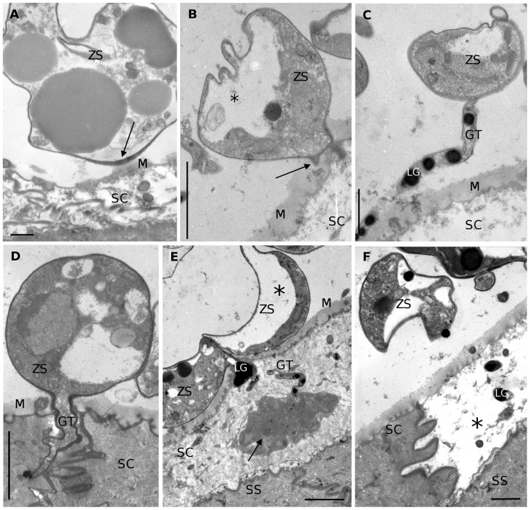
Figure 2. TEM overview of the development of Bd in skin explants of Xenopus laevis.
(A) adhesion of an encysted zoospore (ZS) to the superficial mucus layer (M) on top of the stratum corneum (SC); at the site where adhesion occurs the cell wall of the encysted zoospore is remarkably thickened (arrow); scale bar = 500 nm; (B) initiation of germ tube development (arrow); note the polarisation of the cell cytoplasm (*); scale bar = 2 µm; (C) germ tube (GT) elongating upon the epidermis of X. laevis, with the presence of numerous lipid globules (LG) in the germ tube; scale bar = 1 µm; (D) a growing germ tube protruding the stratum corneum; scale bar = 2 µm; (E) invasion of a host cell resulting in the loss of cell cytoplasm; remnants of the host cell cytoplasm (arrow) are seen at the tip of a protruded germ tube; note the presence of a collapsed sporangium (ZS) due to cell polarisation (*); (SS): stratum spinosum; scale bar = 2 µm; (F) infected epidermal cell with digested cell content (*) alternated by an uninfected normal epidermal cell; note the presence of lipid globules in the infected host cell; scale bar = 1 µm.

