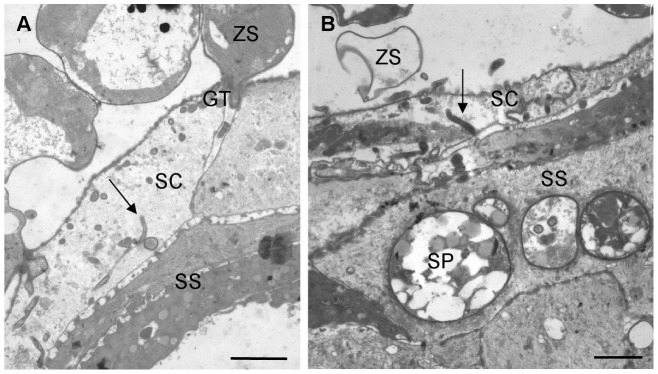
Figure 4. TEM overview of the development of Bd in skin explants of Alytes muletesis and Litoria caerulea.
(A) infected epidermis of A. mulentensis at 1 dpi, with loss of the host cell cytoplasma and the presence of germ tube fragments inside the infected cell in cross and longitudinal section (arrow); scale bar = 2 µm; (B) infected epidermis of L. caerulea at 2 dpi showing colonization of the stratum corneum, loss of the host cell cytoplasm and the presence of germ tube fragments (arrow); intracellular chytrid sporangia are observed in the stratum spinosum; scale bar = 2 µm; GT; germ tube, SC: stratum corneum, SP: sporangium, SS: stratum corneum, ZS: encysted zoospore.

