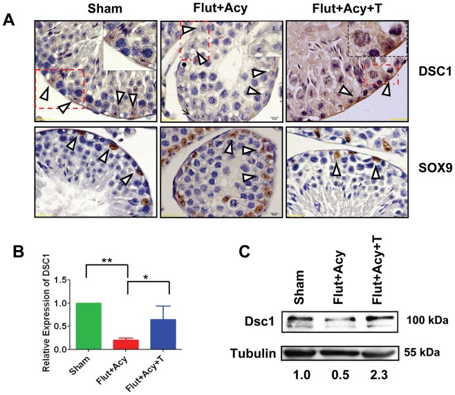
Figure 7. Androgen-dependent expression of Dsc1 in Sertoli cells.
(A) Immunohistochemical analysis on testis sections from Sham, flutamide-acyline (Flut+Acy) and flutamide-acyline testosterone-replacement (Flut+Acy+T) mice, using antibodies against Dsc1 (1∶50) and Sox9 (1∶500). Insets show Sertoli cell-specific expression of Dsc1 in magnified testicular section from Sham, Flut+Acy, and Flut+Acy+T mice. (B) Real-time PCR analysis of Dsc1 transcript levels in total cellular RNA isolated from the purified Sertoli cells from Sham, Flut+Acy, and Flut+Acy+T mice testes. All values are normalized against RNU19 levels. (C) Western blot analysis on total testicular lysates from Sham, Flut+Acy, and Flut+Acy+T mice by using anti-Dsc1 antibody (1∶250). Tubulin was used as a loading control. Values below the gel were quantified using the Image J software. Dsc1 protein level for the control was set to 1. All images were taken at magnification of 600×.

